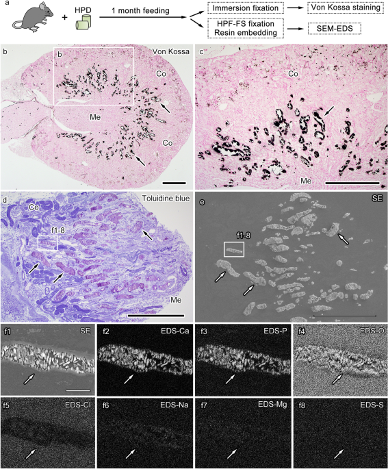
Correlative light and electron microscopic observation of calcium phosphate particles in a mouse kidney formed under a high-phosphate diet
By A Mystery Man Writer
Last updated 16 Sept 2024

Selective affinity of Aln-FITC towards HAp and bone. (a) Fluorescence

Localization of IL‐36α expression induced by phosphate load. Wild‐type

A carbon nanotube tape for serial-section electron microscopy of brain ultrastructure. - Abstract - Europe PMC
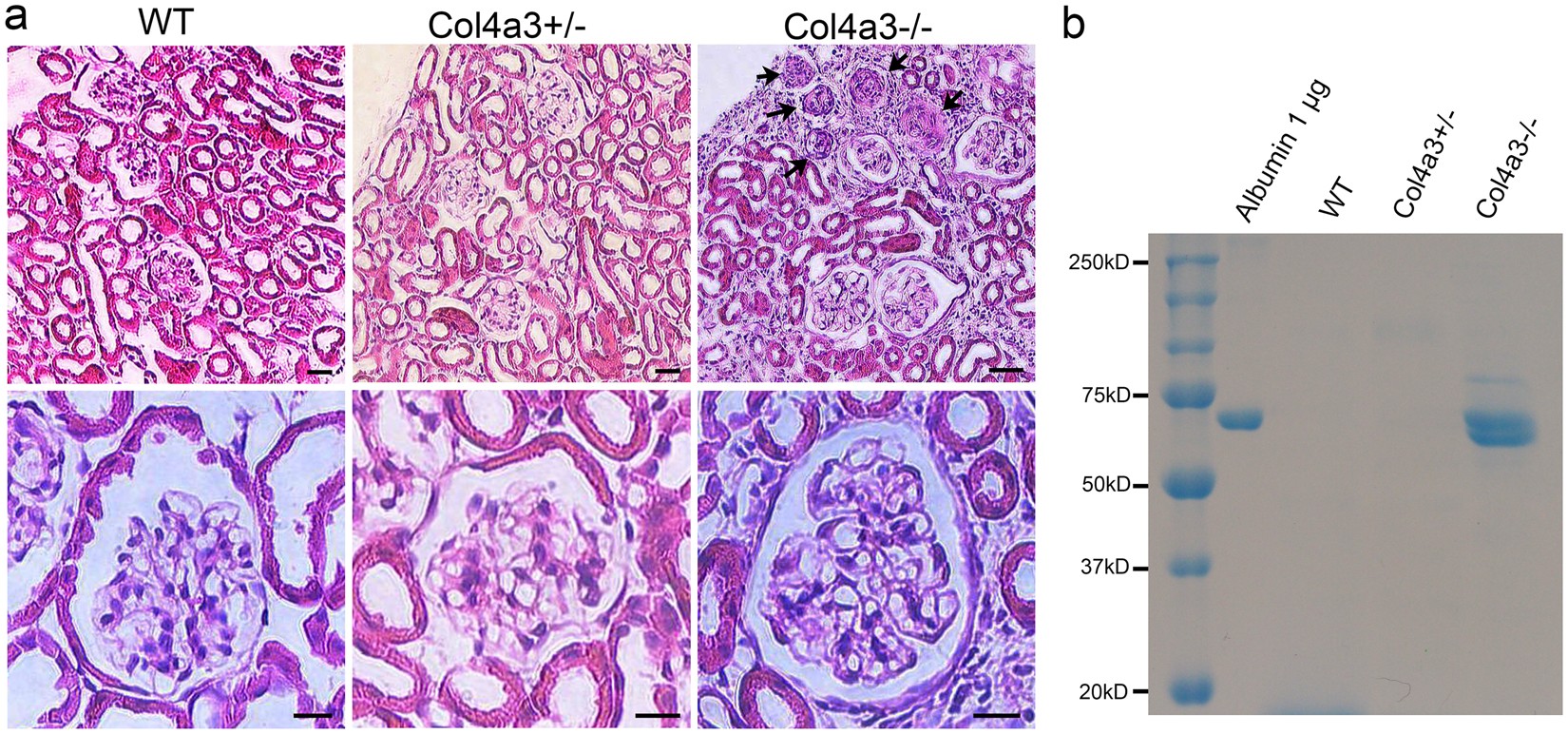
Ultrastructural Characterization of the Glomerulopathy in Alport Mice by Helium Ion Scanning Microscopy (HIM)

Identification of biological components for sialolith formation organized in circular multi-layers

DIVISION OF HISTOLOGY, JICHI MEDICAL UNIVERSITY, SCHOOL OF MEDICINE

CPP in fresh plasma. Total CPP levels were measured in plasma samples

Bisphosphonate binds selectively and quantitatively to fetuin-A
DIVISION OF HISTOLOGY, JICHI MEDICAL UNIVERSITY, SCHOOL OF MEDICINE

Transmission electron microscopic and X-ray absorption fine structure spectroscopic investigation of U repartition and speciation after accumulation in renal cells

PDF) Correlative light and electron microscopic observation of calcium phosphate particles in a mouse kidney formed under a high-phosphate diet

Transmission electron microscopic and X-ray absorption fine structure spectroscopic investigation of U repartition and speciation after accumulation in renal cells

Identification of biological components for sialolith formation organized in circular multi-layers
Recommended for you
 PARIS to LONDON on the incredible Eurostar UNDER THE SEA!14 Jul 2023
 US Factory Building at Record High Delivers Big Boost to Growth - Bloomberg14 Jul 2023
 U.S. shoots down high-altitude object over Alaska - CBS News14 Jul 2023
 15 Best Sunken Living Room Design Ideas for Your Home14 Jul 2023
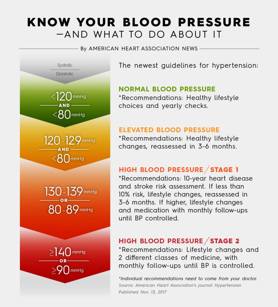 Nearly half of U.S. adults could now be classified with high blood14 Jul 2023
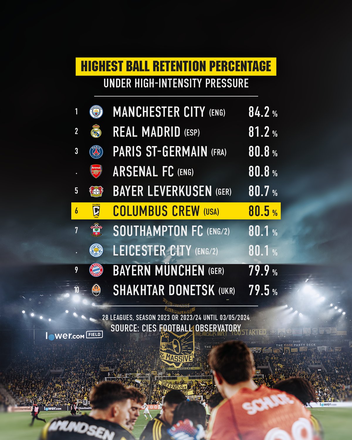
- The Crew (@ColumbusCrew) / X14 Jul 2023
 Chemistry under high pressure14 Jul 2023
 Lower Dauphin High School / Overview14 Jul 2023
 Touché Éclat High Cover Radiant Glow Concealer - YSL Beauty14 Jul 2023
 Deciding Appeal Based On Report By Authority Whose Decision Itself Is Under Challenge Amounts To Violation Of Natural Justice: Patna High Court14 Jul 2023
You may also like
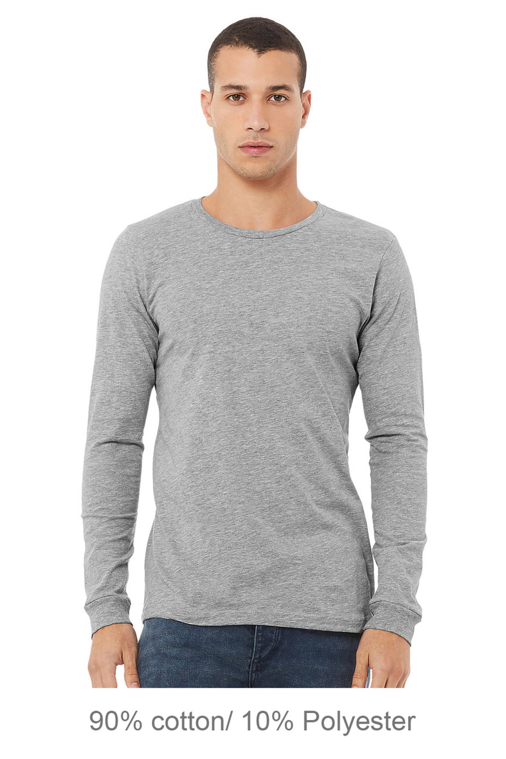 Bella+Canvas 3501CVC Unisex Heather CVC Long Sleeve Tee14 Jul 2023
 Le Mystere Hi Impact Sports Bra 92014 Jul 2023
 PPF & Clear Bra Removal Seattle, Bellevue, Lynnwood14 Jul 2023
 Multi-Surface Wipes - Garden Mint14 Jul 2023
 Calcetines Largos con Cashmere de Mujer - Calzedonia14 Jul 2023
 Old Navy Full-Length Fleece-Lined Leggings for Toddler Girls14 Jul 2023
 UNLEASHIA - 7 Get Loose Glitter Gels14 Jul 2023
 Buy SKIMS White New Vintage Long Sleeve T-shirt - Marble At 2514 Jul 2023
 Pickpocket Proof Vest Women Lapel Loose Blouse Casual Fashion Loose Denim Waist Coat Sleeveless Vest Button Hooded Detachable Coat Space Hood Mid14 Jul 2023
 Rebecca small chest bra A cup AA flat chest gathered comfortable14 Jul 2023