Figure 6 from Femoral Hernia: A Review of the Clinical Anatomy and
By A Mystery Man Writer
Last updated 08 Sept 2024

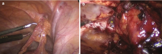
Figure 6. Femoral hernia repair in clean operation. (a) The narrow side of the mesh is sutured to Cooper’s ligament; (b) The mesh is sutured to the iliopubic tract or shelving portion of the inguinal ligament; (c) The posterior wall of the inguinal canal is reinforced, as in Lichtenstein’s repair. - "Femoral Hernia: A Review of the Clinical Anatomy and Surgical Treatment"

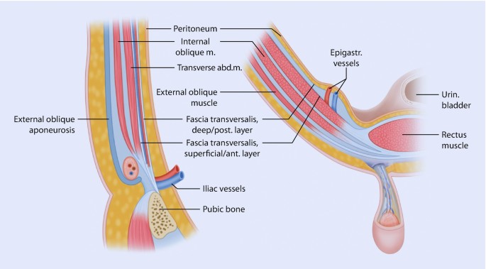
Anatomy of the inguinal and femoral regions. (A) Transversalis fascia

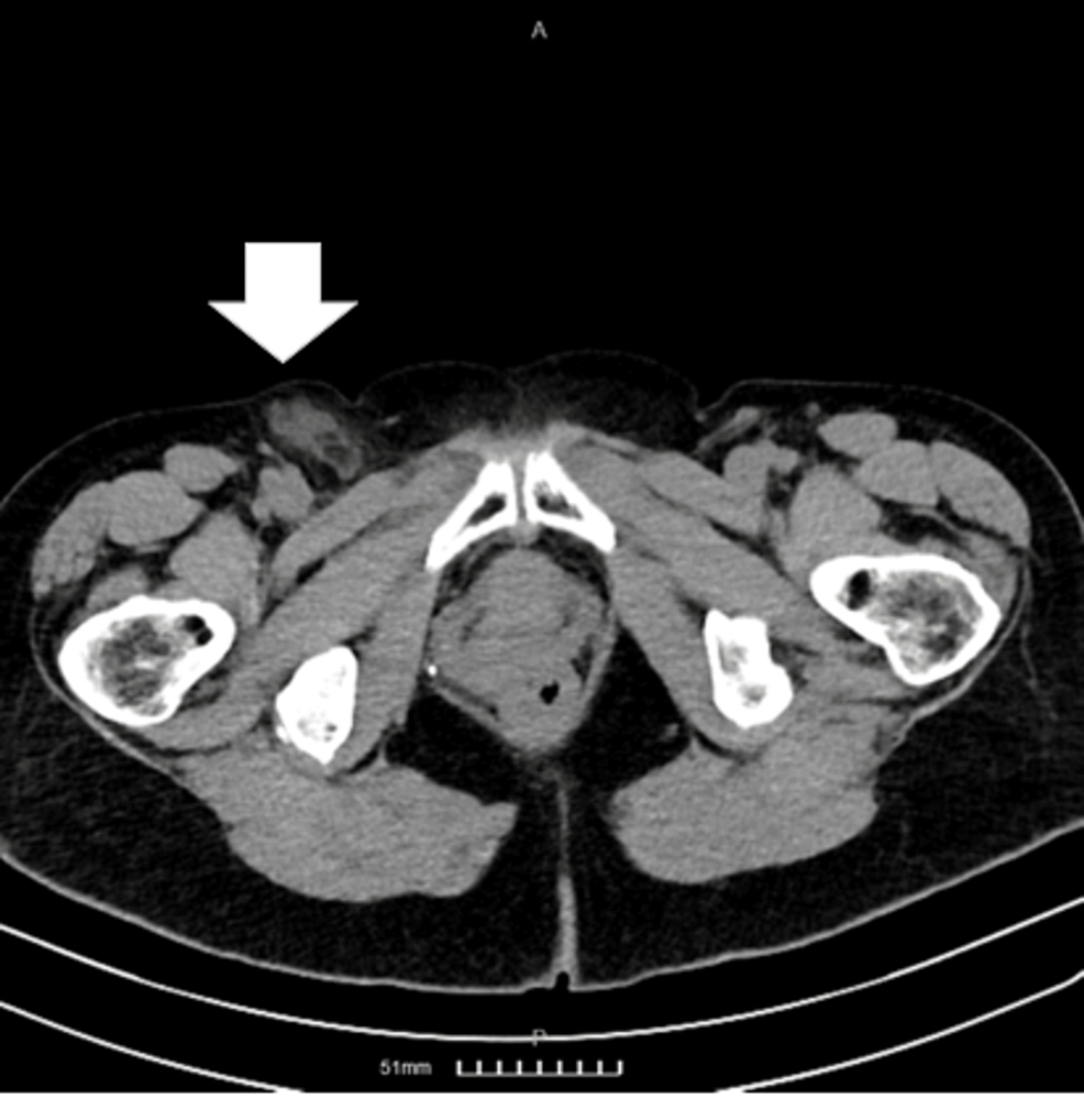
Femoral hernia on herniography, Radiology Case
Clinical Anatomy of the Groin: Posterior Laparoscopic Approach
Cureus, Femoral Hernia Containing a Strangulated Appendix: A Hybrid Approach
Femoral Hernia - A Review of Clinical Anatomy

Femoral Hernia - A Review of Clinical Anatomy

Femoral Hernia: A Review of the Clinical Anatomy and Surgical Treatment

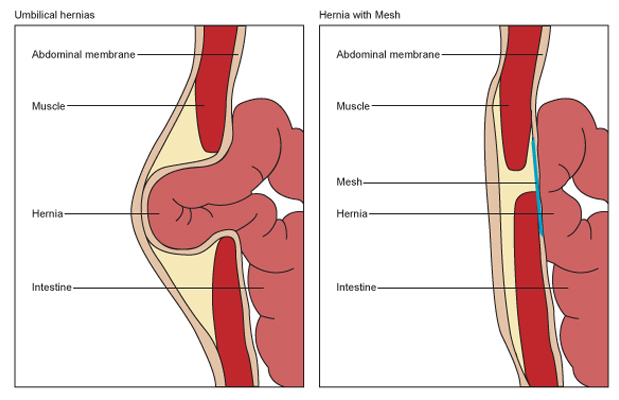
Femoral hernia: Symptoms, pictures, treatments, and more

Clinical Anatomy of the Groin: Posterior Laparoscopic Approach
Femoral Hernia

From inguinal to giant femoral hernia: An unusual postoperative twist - A rare case report - ScienceDirect
Recommended for you
:max_bytes(150000):strip_icc()/137517774-56a9125d3df78cf772a35d63.JPG) Femoral Hernia Surgery: Treatment, Recovery, and More14 Jul 2023
Femoral Hernia Surgery: Treatment, Recovery, and More14 Jul 2023 A: large femoral hernia in a female patient. B: the hernia sac with its14 Jul 2023
 Femoral Hernia Treatment London14 Jul 2023
- A: large femoral hernia in a female patient. B: the hernia sac14 Jul 2023
 Hernia, Inguinal hernia, Femoral hernia14 Jul 2023
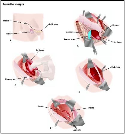 Femoral Hernia Repair - procedure, recovery, blood, pain, complications, adults, time, infection14 Jul 2023
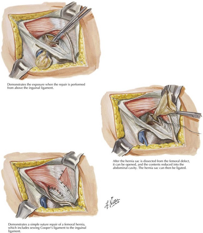 Femoral Hernia Repair14 Jul 2023
 How open femoral hernia surgery is carried out14 Jul 2023
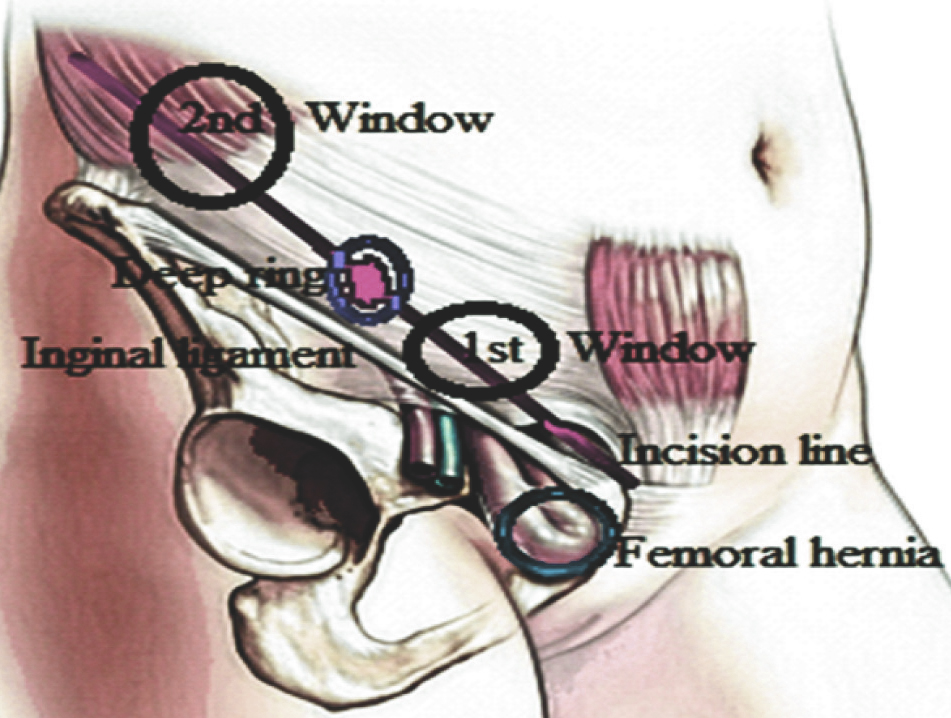 JCDR - Femoral hernia, Gangrenous bowel, Inguinal hernia, One skin incision, Strangulation, Window14 Jul 2023
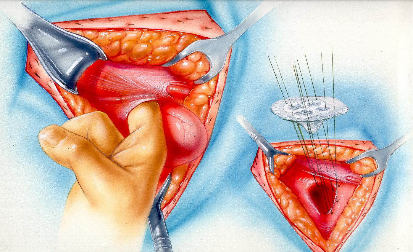 TRABUCCO HERNIA INSTITUTE14 Jul 2023
You may also like
 Fajas Colombianas Women Double Compression Waist Trainer Corset14 Jul 2023
 BARBIE PLAY POUCH TOY STORAGE BAG & MAT – Toyworld Australia14 Jul 2023
 Salmon pink color Color Palette14 Jul 2023
 Jockey® Essentials Women's Seamfree® Eco Plunge Bralette, Wirefree Adjustable Bra, Sizes Small-3XL, 568614 Jul 2023
 Nordic skiing is growing': Local athletes return from OFSAA - Sault Ste. Marie News14 Jul 2023
 ANNYISON Womens Underwear,High Waist Full Coverage Cotton Brief Colorful Panties for Women14 Jul 2023
 Agnes Orinda Plus Size Denim Overall Dress for Women Distressed Suspender Pinafore Dresses with Pockets 1X Dark Blue : Clothing, Shoes & Jewelry14 Jul 2023
 LoriEr Women's Lace Sexy Bra See Through Sheer Unlined Minimizer Full Coverage Underwire Big Breast Bras Black at Women's Clothing store14 Jul 2023
 Vanity Fair Band Bras for Women14 Jul 2023
 Scholl Flight Socks Cotton Feel Size 6.5-9 - Pharmacy & Health from Chemist Connect UK14 Jul 2023